Cardiovascular Ultrasound
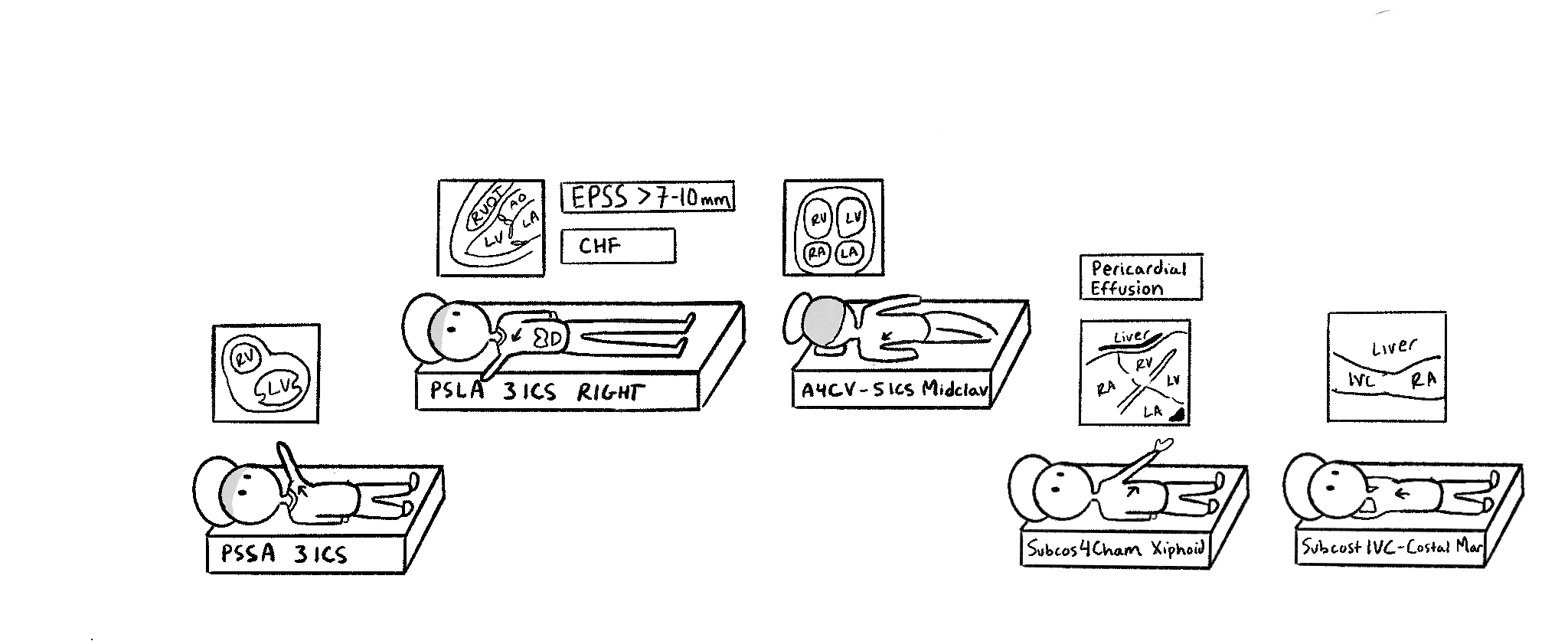
Another day in the ultrasound lab. In this sketch, we added the five major views for ultrasound. Our characters’ arms represent the direction of the ultrasound indicator, except for our third guy, who is lying on his side. For him, his indicator is towards the left axilla. To differentiate Parasternal Long from Short Axis, we made the long axis guy have much longer legs than the short axis guy.
Next, we will look at two clinical cases. One of a heart failure patient, seen with PSLA. And another with pericardial effusion with our Subcostal 4 Chamber View. Notice that the PSLA view has two signs, “CHF” and “EPSS > 7-10 mm” (The distance from the open mitral valve to the septum is called the EPSS. It increases in length due to left ventricle dilation, leading to sluggish filling, and the mitral leaflet does not move as far towards the septum, leading to further distance). On the Subcostal 4-chamber view, our patient has a pericardial effusion. Notice the dark shadows around the heart. Fluid surrounding the heart can make it difficult to pump blood, making it an emergency.