EKG Ischemia
This chapter will review Left bundle branch block and EKG ischemia.

Risk of ischemia is highest with occlusion to the LAD, RCA, then LCX. LAD especially because it covers a large portion of heart muscle responsible for pumping blood into the body, and thus require a lot of oxygen.
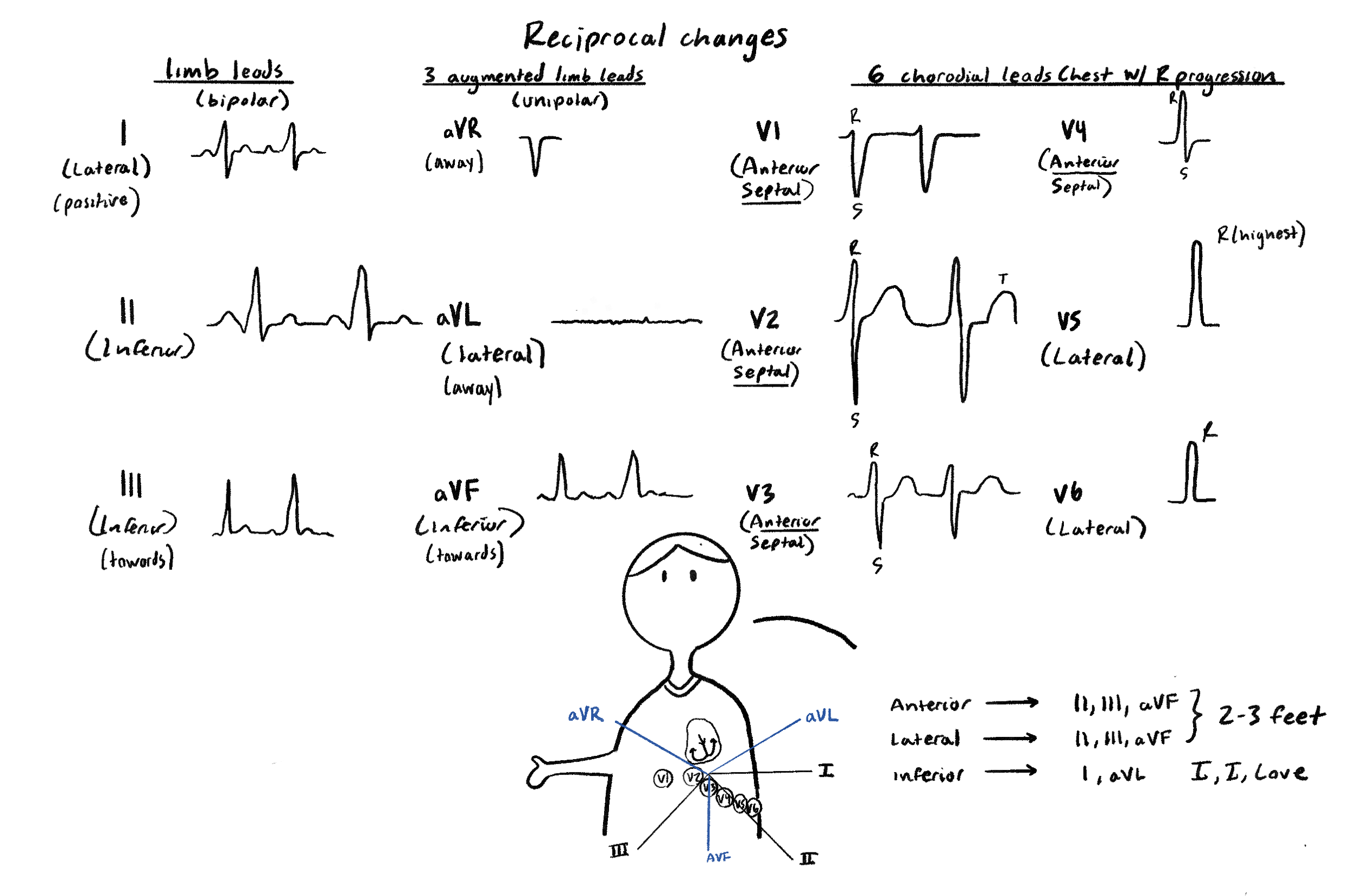
Reciprocal changes just mean that if you have ST elevation on certain contiguous leads, you may expect to see reciprocal signaling on these ones. For example, if there were inferior leads showing ST elevation, we should expect to see ST depression on I, aVL.
A quick way to remember these is (I I Love) and (2-3 Feet)
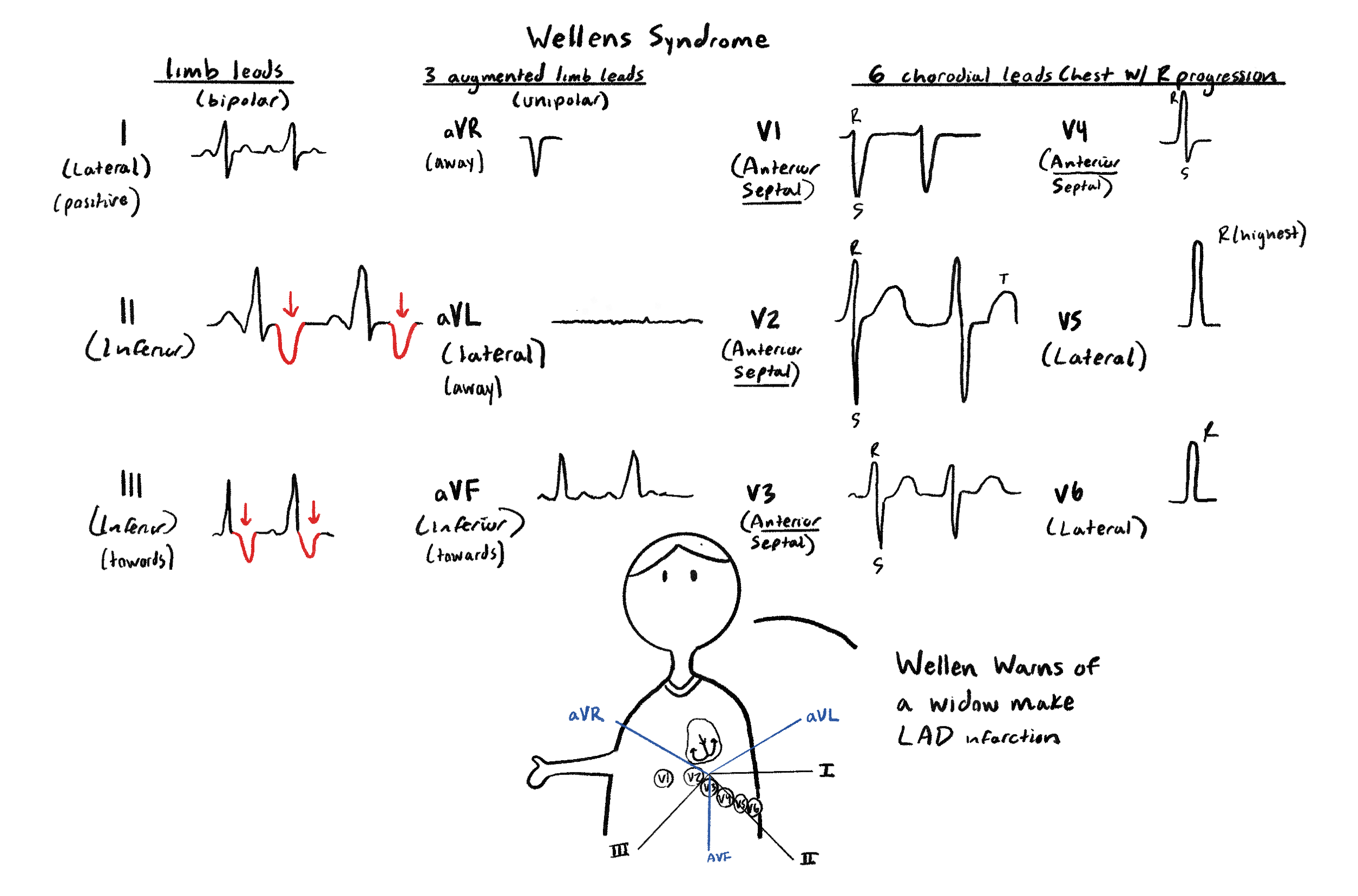
Wellen forms a deep WELL in leads II and III.
Wellen’s Syndrome is lead 2 and 3 with T wave inversion or biphasic, indicating possible risk of LAD occlusion. Patients may not have symptoms at the moment. It is important to notice this sign and know they are at high risk for a myocardial infarction.
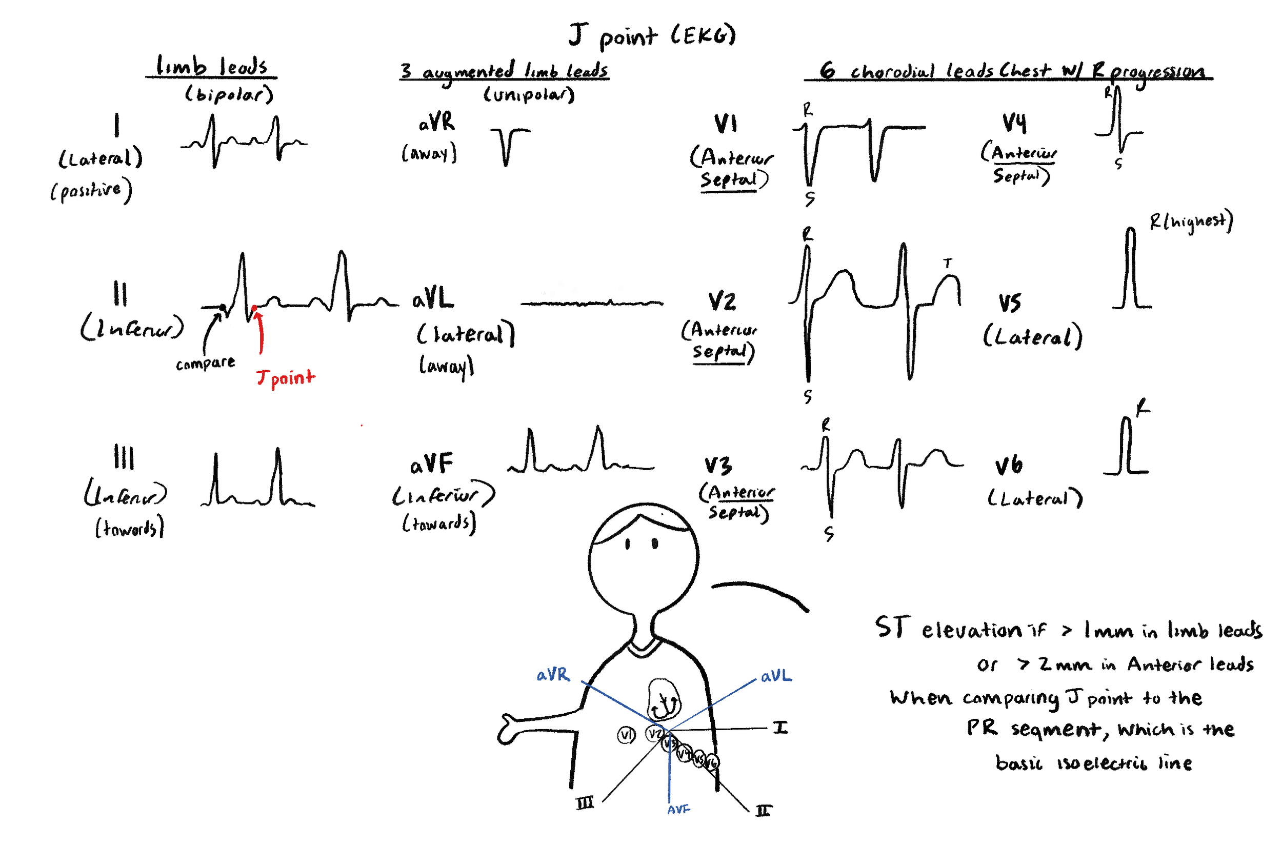
The J point is the “suppose to be normal” beginning isoelectric point of the ST segment. However, it may be elevated or depressed when there is myocardial infarction. As a result, we compare it to the PR segment isoelectric point to see if there truly is an elevation or depression. For elevation, note it has to be > 1 mm in limb leads but > 2 mm in anterior leads
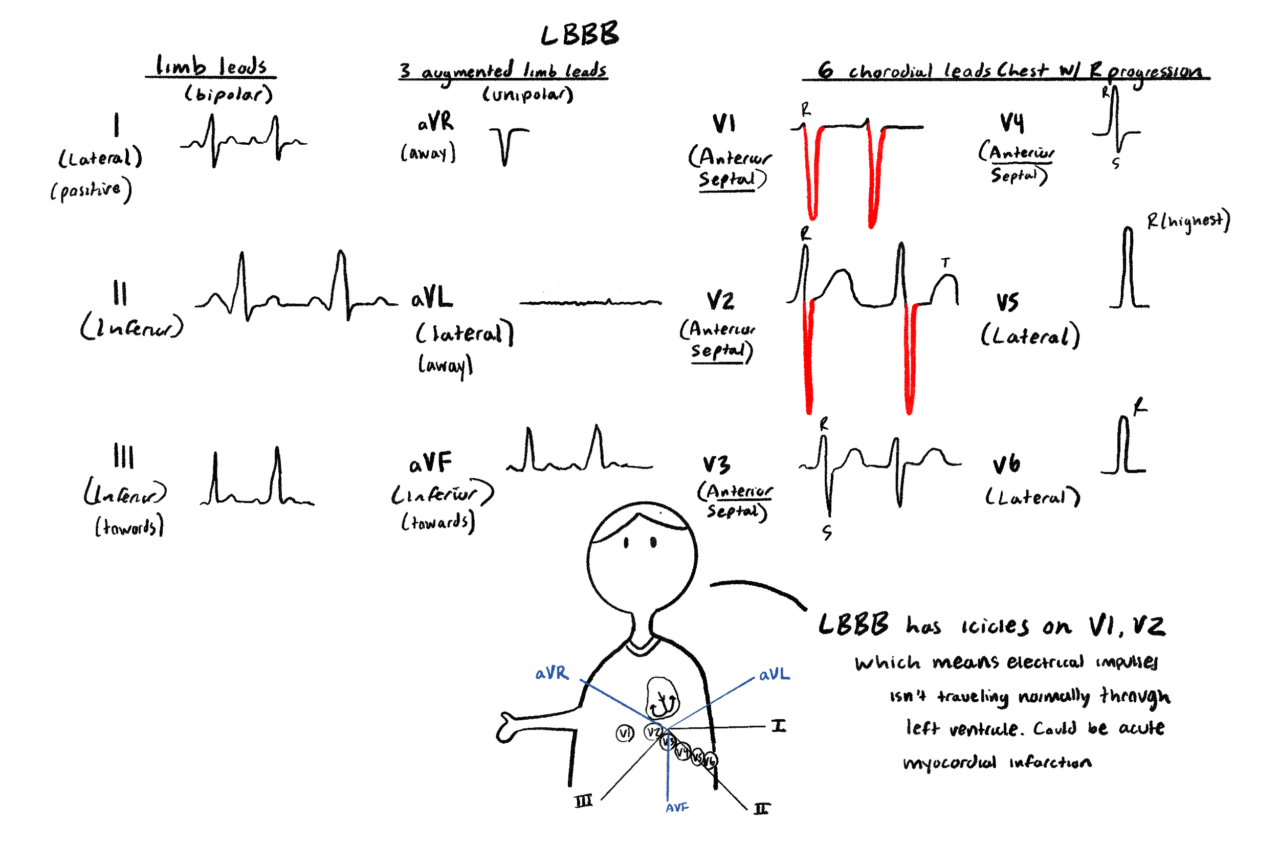
This is a Left Bundle Branch Block, indicating a possible myocardial infarction. LBBB has three Bs, which look like icicles. And you can remember it is on V1 and V2 since V1 + V2 = 3 icicles
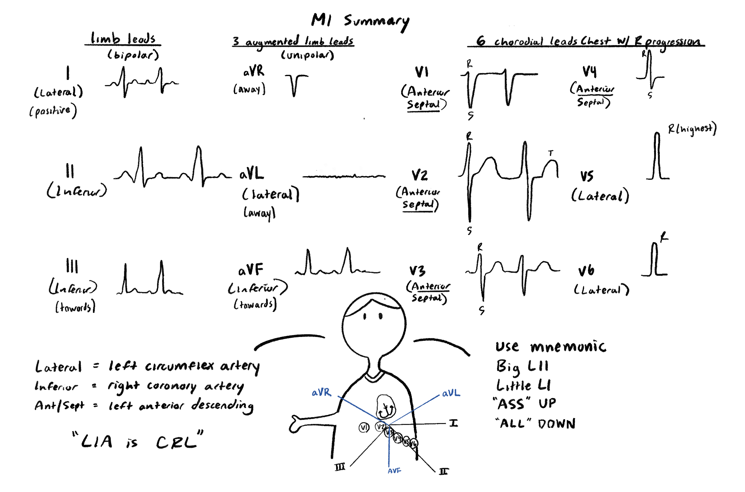
This is a common mnemonic students use for remembering the myocardial infarction localization. The key is to remember that LIA is (CRL) surreal.
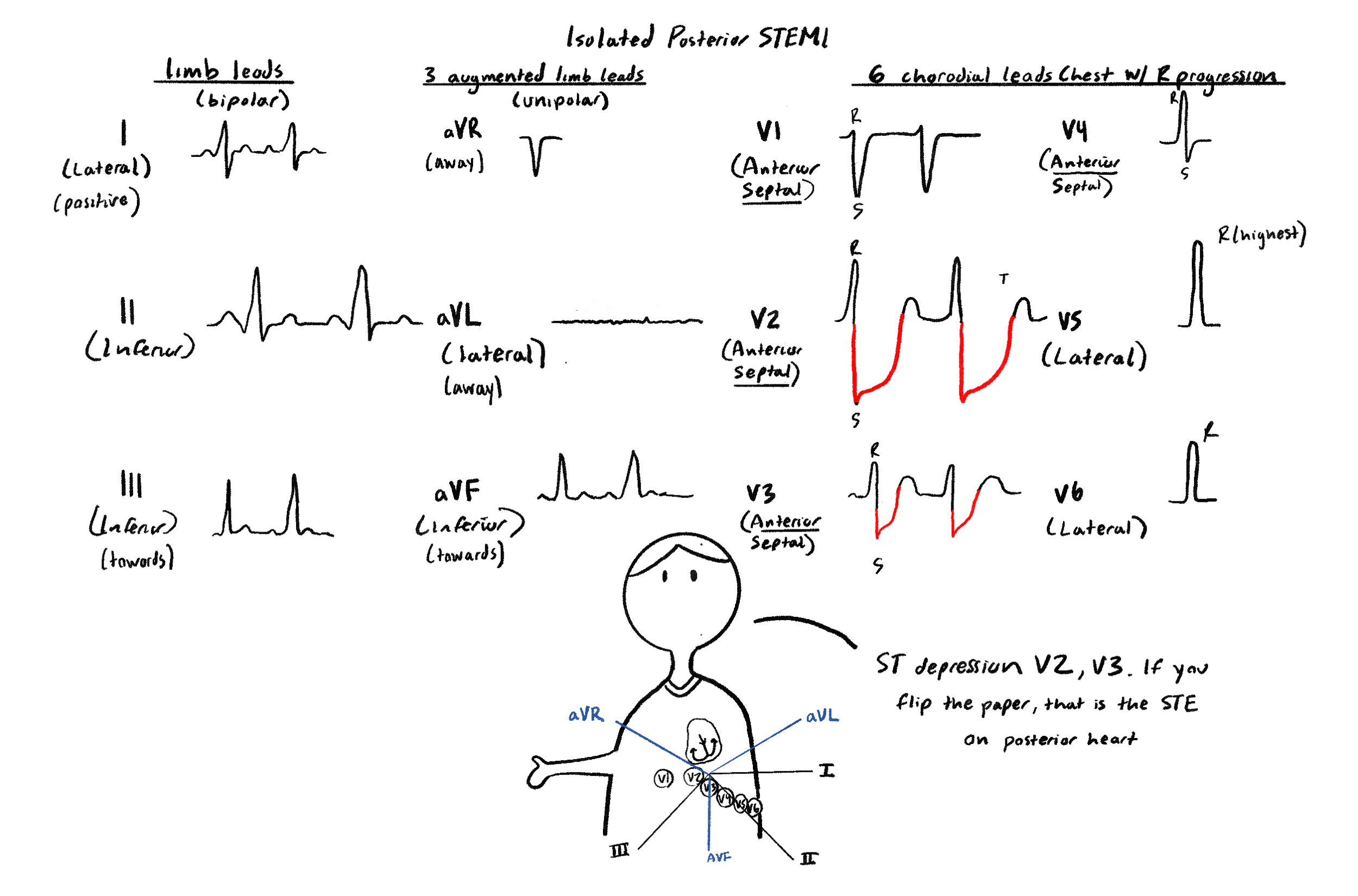
Posterior MI will have V2 and V3 ST depression. Think about posterior being opposite, where you get depression.
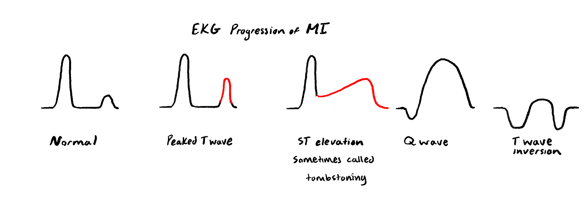
This is EKG progression of MI. Notice that it goes T up, ST ,Q wave,T down. So basically a T sandwich with STQ in the middle, which is alphabetically in order. What do they mean? A peaked T wave indicates acute MI. Then ST elevation means there is transmural damage. “Pathological Q waves” mean irreversible necrosis. And T down waves could mean chronic effects after the MI, which may persist after the event.
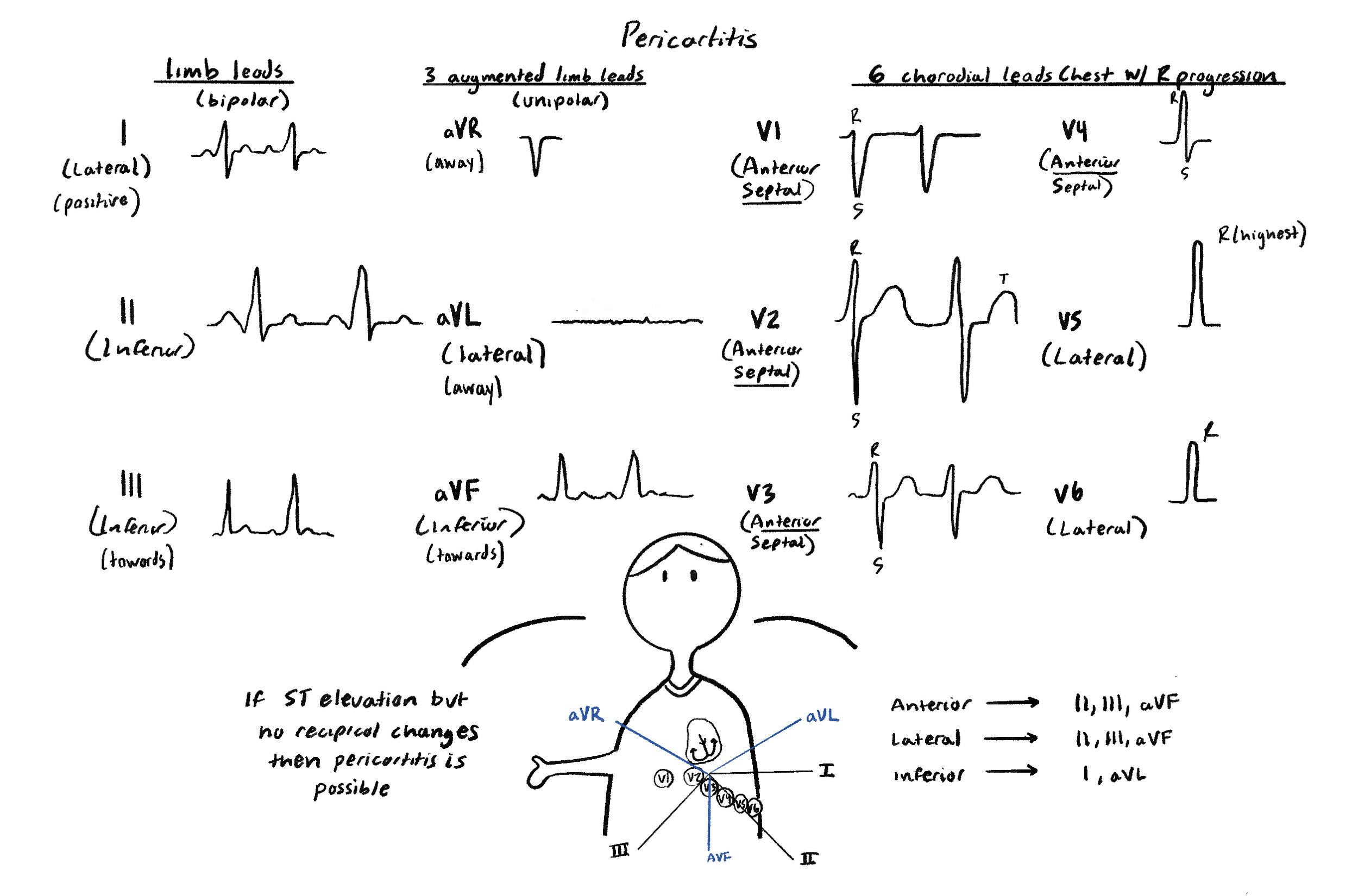
Pericarditis is possible if there is ST elevation but no reciprocal changes in the EKG